
Figure 6
- ID
- ZDB-FIG-210915-93
- Publication
- Schellens et al., 2021 - Zebrafish as a Model to Evaluate a CRISPR/Cas9-Based Exon Excision Approach as a Future Treatment Option for EYS-Associated Retinitis Pigmentosa
- Other Figures
- All Figure Page
- Back to All Figure Page
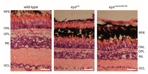
Histological examination of wild-type, eysKO and eysΔexon40-44 zebrafish retinas. Light microscopy of retinal sections from adult zebrafish (15 months post fertilization (mpf)) stained with hematoxylin (purple) and eosin (red). Retinas of the eysΔexon40-44 and eysKO lines are morphologically indistinguishable. In both the eysΔexon40-44 and eysKO retinas, a reduction of the thickness of all retinal layers was observed as compared to wild-type controls. In addition, photoreceptor outer segments seem to be shortened and disorganized in both eysΔexon40-44 and eysKO as compared to wild-type retinas. Scale bar: 20 μm. RPE: retinal pigment epithelium, ONL: outer nuclear layer, OPL: outer plexiform layer, INL: inner nuclear layer, GCL: ganglion cell layer. |
| Fish: | |
|---|---|
| Observed In: | |
| Stage: | Adult |