FIGURE

Figure 6

ID
ZDB-FIG-210606-165
Publication
Rohrer et al., 2021 - Conditional Loss of the Exocyst Component Exoc5 in Retinal Pigment Epithelium (RPE) Results in RPE Dysfunction, Photoreceptor Cell Degeneration, and Decreased Visual Function
Other Figures
All Figure Page
Back to All Figure Page
Figure 6

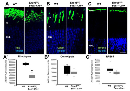
Immunohistochemical analysis of rod and cone photoreceptors in wild-type and conditional Exoc5 knockout mice at 20 weeks of age. Levels and localization of rhodopsin (green, Rho, (A)), red/green cone opsins (green, R/G Opsin, (B)), and RPE65 (green, RPE65, (C)) were assessed using immunohistochemistry in retinal sections of 20-weeks-old mice to identify alterations in rod and cone outer segments, as well as retinal pigment epithelium (RPE). Only moderate differences in staining were observed. Scale bars = 50 μm (AC). Image J was used to quantify rhodopsin (A’) and cone opsin (B’) fluorescence in photoreceptors, and RPE65 (C’) in RPE of WT and Exoc5fl/fl;Best1-Cre+ mice. OS, outer segments; IS, inner segments; ONL, outer nuclear layer, INL, inner nuclear layer; RPE, retinal pigmented epithelium. ** p < 0.05 (WT vs. Exoc5fl/fl;Best1-Cre+ mutants).

Expression Data

Expression Detail
Antibody Labeling
Phenotype Data

Phenotype Detail
Acknowledgments
This image is the copyrighted work of the attributed author or publisher, and ZFIN has permission only to display this image to its users. Additional permissions should be obtained from the applicable author or publisher of the image. Full text @ Int. J. Mol. Sci.