
Figure 1
- ID
- ZDB-FIG-210508-18
- Publication
- Feitosa et al., 2021 - Brazilian silverside, Atherinella brasiliensis (Quoy & Gaimard,1825) embryos as a test-species for marine fish ecotoxicological tests
- Other Figures
- All Figure Page
- Back to All Figure Page
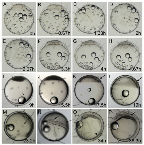
(A, B, F–M, O, and P) lateral view and (C, D, E, and N) dorsal view. (A) 1-cell stage, considered 0 h from fertilization until first cleavage; (B) 2-cell stage, 0,67 hpf; (C) 4-cell stage, 1.33 h; (D) 8-cell stage, 2 h; (E) 16-cell stage, 2.67 h; (F) 32-cell stage, 3.3 h; (G) 64-cell stage, 4 h; (H) 128-cell stage; 4.67 h; (I) oblong stage; 9 h; (J) sphere stage, 15.5 h; (K) 25% epiboly, 17.5 h the embryo is on the right side (arrow); (L) 40% epiboly, 19 h embryo is on the right side (arrow); (M) 90% epiboly, 23.2 h; (N) optic vesicles forming at 90% epiboly stage (arrow); (O) 14-somite stage embryo, at 34 h showing Kupffer’s vesicle (arrowhead); (P) 18 somite stage, 38.3 h, the otic vesicle is apparent (arrow) (Scale—0.5 mm) |