|
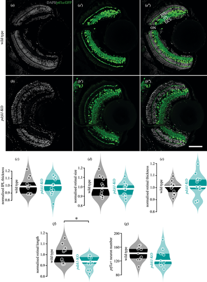
Transgenic labeling of retinal layers and quantification of retinal morphology for wild-type and pdzk1-KO larvae. (a–b″) Inhibitory neurons, including horizontal and amacrine cells, and their projections into the outer and inner plexiform layers were labeled by transgenic construct Tg(ptf1a:GFP) in green, revealing distinct retinal layers in wild-type and pdzk1-KO retinas. Nuclei stained with DAPI are shown in gray. Scale bar: 50 µm. Normalized (c) IPL thickness, (d) retinal size, (e) retinal thickness, (f) retinal length, and (g) ptf1a positive (ptf1a+) neuron number were quantified. Points are data from individual retinas. White lines represent medians, and dark bands indicate interquartile ranges. For all analyses, unpaired t-tests were performed with 12 and 13 retinas for wild-type and pdzk1-KO groups, respectively. *P < 0.05. INL, inner nuclear layer; IPL, inner plexiform layer; GCL, ganglion cell layer; ON, optic nerve.
|