|
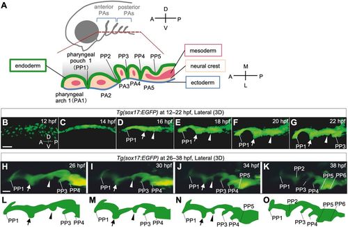
Time-lapse observations of pharyngeal endoderm during PP segmentation in Tg(sox17:EGFP) zebrafish embryos. (A) Schematic of the bilateral arrangement of PAs in the ventral region of the head. (B-K) Time-lapse analysis of the pharyngeal endoderm of Tg(sox17:EGFP) zebrafish from 12 hpf to 22 hpf (B-G, Movie 1) and from 26 hpf to 38 hpf (H-K, Movie 2). Rostral (arrows) and caudal (arrowheads) bulges appeared posterior to PP1 and gradually fused to form PP2. (L-O) Schematic illustrations of the shape of the lateral pharyngeal endoderm in H-K, respectively. A, anterior; D, dorsal; L, lateral; M, medial; P, posterior; PA1-5, the first to fifth pharyngeal arch; PP1-6, the first to sixth pharyngeal pouches; V, ventral. Scale bars: 50 μm (B) and 20 μm (H).
|