Figure 4

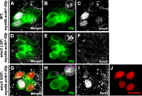
Localization of Emx2 expression in wild-type (WT), loss of function (LOF), and gain of function (GOF) neuromasts at 2.0 dpf.

(A–C) Only hair cells (HCs) (B, Gfp) in the anterior neuromast of WT are positive for anti-Emx2 staining (C). Inset in B shows the bidirectional hair bundle orientation pattern. (D–F) No HCs (E) are positive for anti-Emx2 staining (F) in emx2 LOF neuromast and all hair bundles are oriented in P→A direction (E). (G–J) An emx2 GOF neuromast. (G) Merged image of Gfp (H), anti-Emx2 (I), and mCherry (J) expression. Anti-Emx2 staining only detected endogenous Emx2 in the anterior HCs (I), whereas exogenous Emx2 expression as indicated by mCherry expression is present in all HCs (J). All hair bundles are oriented in A→P direction and two immature HCs show the kinocilium located in the center (magenta arrows).

Expression Data

Expression Detail
Antibody Labeling
Phenotype Data
Fish:
Observed In:
Stage: Long-pec

Phenotype Detail
Acknowledgments
This image is the copyrighted work of the attributed author or publisher, and ZFIN has permission only to display this image to its users. Additional permissions should be obtained from the applicable author or publisher of the image. Full text @ Elife