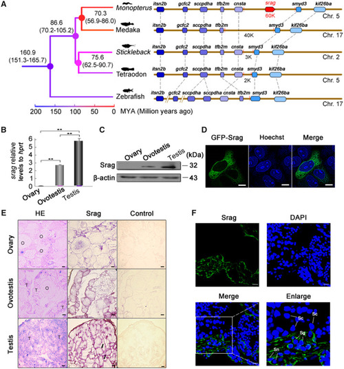
Identification and expression of srag in testis. (A) Synteny analysis. New gene srag in the genome of Monopterus has not any homologous genomic region in other teleost species. srag is a new gene on chromosome 5 of Monopterus; other genes on the chromosome are syntenic and conserved between Monopterus and other fishes (medaka, stickleback, tetraodon, and zebrafish). The number on the nodes indicates the time ranges of divergence from the present (Ma). (B) Quantitative real-time PCR of srag in gonads of Monopterus. Total RNA samples were isolated from ovary, ovotestis, and testis of adult individuals. srag mRNA expression was upregulated from ovary to testis. hprt was used as an internal control. (C) Western blot analysis showed Srag protein levels in different gonads using the anti-Srag antibody. β-Actin was used as an internal control. (D) Srag was expressed in the cytoplasm of HeLa cells after transfection. Images were captured using confocal microscopy. The nuclei were revealed using Hoechst fluorescence (blue). Scale bar: 5 μm. (E) Expression analysis of srag by mRNA in situ hybridization on gonad sections in adult individuals. Positive signals were observed in peripheral region of seminiferous epithelium (arrows). H&E staining indicates tissue structure of gonads. Labeled sense-strand DNA was used as a control. Scale bar: 50 μm. (F) Immunofluorescent localization of Srag protein in gonads using anti-Srag antibody followed by FITC-conjugated ImmunoPure goat anti-rabbit IgG (green). Srag was expressed in the cytoplasm of Sertoli cells (Sn) and spermatogonia (Sg) in testis. The nuclei were stained by DAPI (blue). Images were captured using confocal microscopy. The enlarged images originated from the regions with white squares. Scale bar: 10 μm.
|
