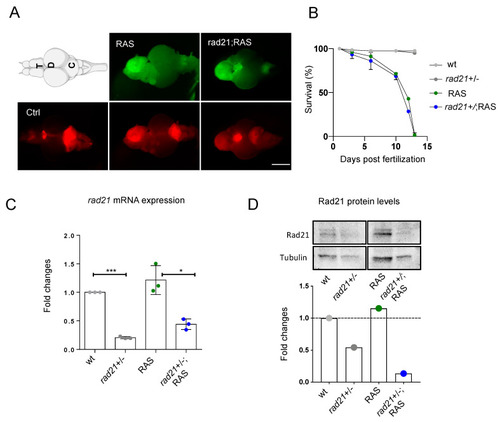
Rad21 heterozygous fish develop brain tumors with the same frequency and location than wt. (A) Representative images of zebrafish RAS and rad21;RAS brains used for this study. The expression of UAS:eGFP-HRASV12 was induced in a population of brain progenitor cells using the driver line zic:GAL4 (visualized through mCherry expression, bottom panel). Tumor formation is visualized through eGFP expression in 1-month-old fish (upper panel). The location of tumors appears to be similar in both RAS and rad21;RAS fish. Scale bar: 0.5 mm. T: telencephalon; OT: optic tectum; C: cerebellum. (B) Survival curve of RAS and rad21;RAS shows a very low survival rate (approx. 1%) for both genotypes n = 2 in control groups (gray dots) and n = 4 in tumor groups (blue and green dots). (C) Expression of zebrafish rad21a mRNA in controls and brain tumors measured by RT-qPCR. Values were normalized to rps11 mRNA levels; n = 3 in all groups; *** p < 0.001; * p < 0.05. (D) Rad21 protein levels in Western blot and relative quantification. Rad21 levels are reduced in control (2nd lane) and tumor brains (4th lane) from rad21ahi2529Tg/+ fish compared to the wild type (fold changes). Values were normalized to tubulin levels.
|
