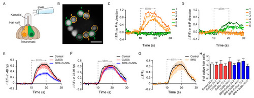
Calcium imaging reveals the function of a single neuromast. (A) The schematic diagram shows a glass micropipette filled with fluid located about 100 μm away from the top of kinocilia to stimulate the neuromast. The orange and green hair cells represent different polarities. (B) When stimulated by the flow, only a portion of hair cells responded in this focal plane (circled cells), and some were far from this focal plane (dashed circled cells). The No. 2, 4, and 6 active hair cells (orange circles) only responded to the flow in the P–A direction (C). At the same time, the No. 1, 3, and 5 active hair cells (green circles) only responded to the flow in the A–P direction (D). Scale bar in (B) represents 10 μm. (E) The relative fluorescence intensity change (ΔF/F0) of the BRS+CuSO4 group was significantly lower than that of the CuSO4 group in the Early Stage of regeneration (within 48 hpi) (p < 0.001). (F) ΔF/F0 of the BRS+CuSO4 group was not significantly different from that of the control group or CuSO4 group in the Late Stage of regeneration (72–96 hpi). (G) There was no difference in ΔF/F0 between the BRS group and the control group. (H) During the regeneration process, the numbers of active hair cells per neuromast in the CuSO4 and BRS+CuSO4 groups were basically the same and did not increase with the total number of regenerated hair cells. For (E–H), comparisons were performed using one-way ANOVA, with Tukey’s multiple comparisons test.
|
