
Fig. S3
- ID
- ZDB-FIG-200730-37
- Publication
- Chang et al., 2020 - Functionally distinct Purkinje cell types show temporal precision in encoding locomotion
- Other Figures
- All Figure Page
- Back to All Figure Page
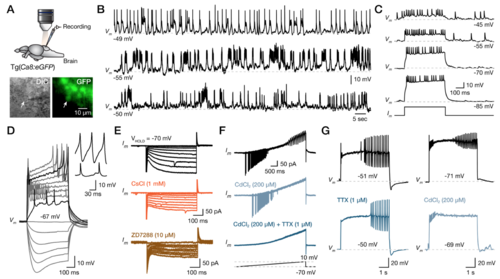
Common physiological properties of the Purkinje cells. (A) Ex-vivo setup of an isolated intact brain from the Tg(Ca8:eGFP) line allows whole-cell patch-clamp recordings of Purkinje cells. Arrows indicate the recorded cell. (B) Sample traces showing the intense and variable spontaneous activity of three different Purkinje cells recorded in the adult zebrafish cerebellum. (C) Application of a bias hyperpolarization current eliminates the spontaneous activity without affecting the Purkinje cell firing pattern induced by a depolarized current step injection. (D) Step current injections reveal the common Purkinje cell properties (sag potential, sodium spikes, calcium spikes). Magnification of the calcium-based (top) and sodium-based (bottom) spikes showing noticeable differences in the amplitude and duration. (E) Voltage-clamp recordings show inward current reduced by Ih blockers (CsCl, ZD7288). (F) Voltage-clamp ramp recordings show sodium (blocked by TTX) and calcium (blocked by CdCl2) spikes. (G) Current-clamp recordings show the elimination of the sodium spikes after the application of TTX and the calcium spikes with CdCl2. CdCl2, calcium chloride; CsCl, cesium chloride; DIC, differential interference contrast; GFP, green fluorescent protein; TTX, tetrodotoxin. |