|
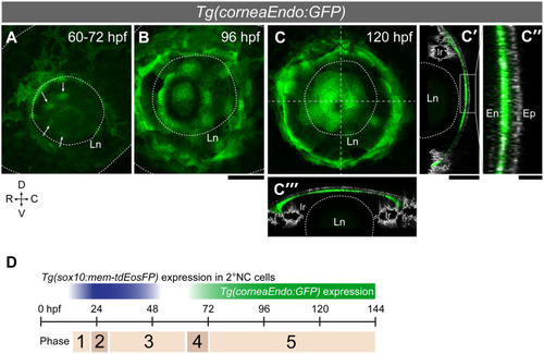
Endothelial cells emerge in a concentric ring pattern in the nascent cornea.(A-C”) The Tg(corneaEndo:GFP) line shows the first corneal endothelium cells (green) as a patch of cells over the lens (arrows in A, 60–72 hpf) that later spread across the entire inner surface of the cornea (B: 96 hpf; C:120 hpf). (C’-C”‘) Transverse (C’) and horizontal (C”‘) optical sections along the stippled lines in C, merged onto pictures from reflection imaging (grey channel) which highlight the corneal epithelium (Ep; note: the strong lateral signals are iridophores). (C”) Magnified view of the central cornea region indicated by a rectangle in C’. En: corneal endothelium; Ln: lens; Ir: iris; Scale bars: 50 μm (A-C, C’ and C”‘); 10 μm (C”). (D) The AS development is classified into five phases. Phase 1: 2°NC cell cluster formation (18–22 hpf); Phase 2: 2°NC cluster dissolves and migrate into the eye (22–28 hpf); Phase 3: NC cells differentiate into ocular mesenchymal cells (28–60 hpf); Phase 4: First corneal endothelial cells emerge over the lens (60–72 hpf); Phase 5: Corneal endothelium fully covers the eye (>72 hpf).
|