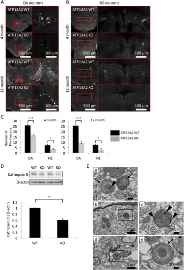
Neuropathology of Atp13a2 deficient zebrafish. (A) Axial sections of the posterior tuberculum containing dopaminergic (DA) neurons of zebrafish brain at 4 and 12 months. Right figures in each section are the enlarged images of the left figures. (B) Axial sections of the locus coeruleus containing norepinephrine (NE) neurons of zebrafish brain at 4 and 12 months. Right figures in each section are enlarged images of the left figures. (C) The number of TH + neurons in the posterior tuberculum and the locus coeruleus. (D) The amount of cathepsin D protein in zebrafish brain. WT indicates wild type and KO indicates Atp13a2 deficient zebrafish. (E) Transmission electron microscopy analysis of the middle diencephalon samples from Atp13a2 deficient zebrafish. Atp13a2 deficiency induces lysosome-like bodies that contain granular deposits (arrowheads) and fingerprint-like structures (arrows). Right figures in each section are the enlarged images of the left figures. Scale bars are indicated in A, B and E. *p < 0.05 and ***p < 0.001.
|
