|
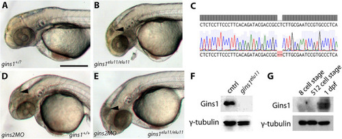
Gins1 deficient and Gins1, Gins2 double-deficient embryos show comparable cell death phenotypes in eyes and tecta. (A,B) The gins1elu11 frameshift allele created through genome editing shows retinal and tectal apoptosis at 2 dpf, similar to other CMG mutants. (C) Sanger sequencing of gins1elu11/elu11 embryos shows the presence of the homozygous c.250_251delCT mutation. (D,E) Phenotype of gins1 mutant embryos and their siblings injected with 100 μM gins2MO. (F) Western blot analysis of protein lysates from 2 dpf control and gins1elu11/elu11 embryos probed with Gins1 and γ-tubulin antibodies. (G) Western blot analysis of protein lysates from wildtype embryos at the indicated stages probed with Gins1 and γ-tubulin antibodies. Scale bar: 250 μm.
|