|
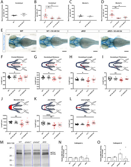
Craniofacial deficits are rescued by Od-mediated inhibition of cathepsin K. (A-D) Mean fluorescence intensity of Col2 immunostaining in the ceratohyal (WT in A, dKO in B) and Meckel's cartilage (WT in C, dKO in D) of 4 dpf larvae with and without Od treatment. (E) Representative images of larvae stained with Alcian Blue. Scale bars: 200 µm. (F-L) Craniofacial morphological measurements at 4 dpf. The measured parameters are highlighted in red in the schematics. (M) In-gel analyses of BMV109, showing cathepsin activities in WT and pheta1/2 mutants at 4 dpf. Blue lines indicate the lane boundaries. (N,O) Quantitation of the cathepsin K and cathepsin L bands from four experiments. Error=s.e.m. ch, ceratohyal; Ctsk, cathepsin K; Ctsl, cathepsin L; m, Meckel's cartilage. *P<0.05, **P<0.01, ****P<0.0001.
|