|
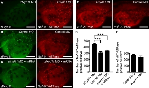
Effects of zFxyd11 knockdown. (A–C) Immunofluorescence staining was performed on zfxyd11-MO-injected (A), control-MO-injected (B), and zfxyd11-MO with zfxyd11a mRNA-injected larvae (C) with anti-zFxyd11 antiserum (green), along with anti–Na+–K+-ATPase antiserum (red). No signal for zFxyd11 was detected in zfxyd11 morphants (A). (D) The number of Na+–K+-ATPase–positive cells in the skin was counted and is represented as the mean ± SEM. Significant differences were evaluated by one-way ANOVA, Tukey's multiple comparison test (***p < 0.001, n = 7). (E) Immunofluorescence staining was performed on zfxyd11-MO-injected (left panel), control-MO-injected larvae (right panel) with anti–vH+-ATPase antiserum. (F) The number of vH+-ATPase–positive cells in the skin was counted and is represented as the mean ± SEM. The number of vH+-ATPase-positive cells was not affected by zFxyd11 knockdown (n = 7).
|