|
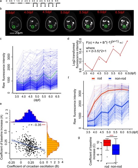
Developmental dynamics of <italic>nr1d1</italic>:VNP expression.(a) Experimental design to examine the developmental dynamics of nr1d1:VNP expression. (b) Fluorescence images illustrate the tracing of one nr1d1:VNP cell during development from 3.5 dpf to 6.5 dpf. This cell was highlighted in green in Fig 3C. (c) Raw fluorescence intensity of the traced cells (six fish). (d) Regression model used to fit the fluorescence intensity (the cell is illustrated in Fig 3B and highlighted in Fig 3C). The dots represent the fluorescence intensity, while the line represents the fitting curve. (e) Scatterplot demonstrated the relationship between coefficients of developmental effect (A) and circadian oscillation (B). The histograms in orange and blue showed the distribution of coefficients of developmental effect (A) and the distribution of coefficients of circadian oscillations (B), respectively. ***Pearson’s correlation P < 0.001. (f) Raw fluorescence intensity of rod-like cells and non-rod-like cells (six fish). Each thin line represents one cell and each dot represents raw fluorescence intensity. The thick lines represent the loess-smoothed curves for all rod-like cells in red and non-rod-like cells in blue, respectively. The shaded areas show the 95% confidence level of the smoothed curve. (g) A comparison of baseline expression (C) between rod-like and non-rod-like cells. Two-tailed Student t test was applied to calculate the levels of significance between the two types of cells. **P < 0.01. The numerical values for panels c–g were in S1 Data. dpf, days postfertilization.
|