
Figure 4—figure supplement 1—source data 1.
- ID
- ZDB-FIG-200220-47
- Publication
- He et al., 2020 - In vivo single-cell lineage tracing in zebrafish using high-resolution infrared laser-mediated gene induction microscopy
- Other Figures
- All Figure Page
- Back to All Figure Page
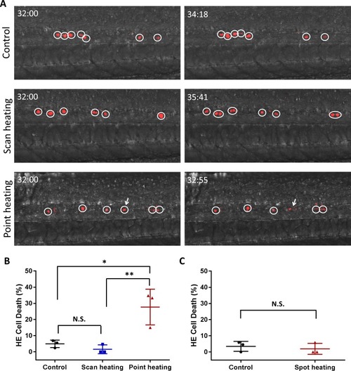
Assessment of HE cell damage caused by IR-LEGO heat shock.(A) Representative time-lapse images of the control embryos (the first row), the scanning heat-shocked embryos (the second row), and the point heat-shocked embryos (the third row). Circles depict the individual converted HEs in the PBI region (dorsal wall on the bottom). Number at the top left corner in each image indicates the developmental stage of the embryos (hh:mm post fertilization). One of the point heat-shock HE (white arrow) burst into fragment, while the HEs in control embryos and scanning heat-shocked keep viability during time-lapse imaging. (B) Quantification of the HE cell death in the control embryos (three independent experiments, in which the number of dead cell/total cell are 1/18, 1/42 and 2/29, respectively. Totally 4 of 89 cells died.), the scanning heat-shocked embryos (three independent experiments, in which the number of dead cell/total cell are 1/21, 0/34 and 0/23, respectively. Totally 1 of 78 cells died.), and the point heat-shocked embryos (three independent experiments, in which the number of dead cell/total cell are 8/23, 9/27 and 3/20, respectively. Totally 20 of 70 cells died.). Statistical analysis indicates that the percentage of HE cell death in the scanning heat-shocked embryos has no difference from the control group, while heat shock fixed on a single point result in significantly more cell death. The percentages of cell death are shown in terms of mean ± standard deviation. One-way ANOVA was used for significance test. *P < 0.05; **P < 0.01. (C) Cell death test on the old IR-LEGO system used for bulk labeling previously. The IR laser beam was loosely focused by a low-NA lens and heat shocked the embryos on each spot for 2 minutes. Statistical analysis shows that the single spot heat shock used for bulk labeling would not cause cell damage. |