
Fig 2
- ID
- ZDB-FIG-200220-23
- Publication
- Messerli et al., 2020 - Adaptation mechanism of the adult zebrafish respiratory organ to endurance training
- Other Figures
- All Figure Page
- Back to All Figure Page
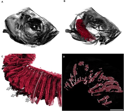
A: Fish head. The diameter of the whole eye (centre marked with a white asterisk) is approximately 0.83 mm. B: The delineated gills in red are shown inside the head of the fish, operculum removed, only right arches of the gills are shown. The gill arches lie within the branchial chamber. In this image, primary filaments are mainly pointing to the left of the image (back of fish). C: Detailed view of gills. Secondary filaments are seen as leaf-like structures attached to the primary filaments. The semitransparent grey line marks one primary filament. Arrows mark the tips of four secondary filaments. D: Two-dimensional view of the gills, e.g. one slice of the tomographic data set where all three-dimensional measurements were based on. The red overlay denotes the estimation of the hull of the gill organ. The filling factor of the gills shown in the right panel of |