FIGURE

Fig. 3

ID
ZDB-FIG-200124-54
Publication
Toms et al., 2019 - Missense variants in the conserved transmembrane M2 protein domain of KCNJ13 associated with retinovascular changes in humans and zebrafish
Other Figures
All Figure Page
Back to All Figure Page
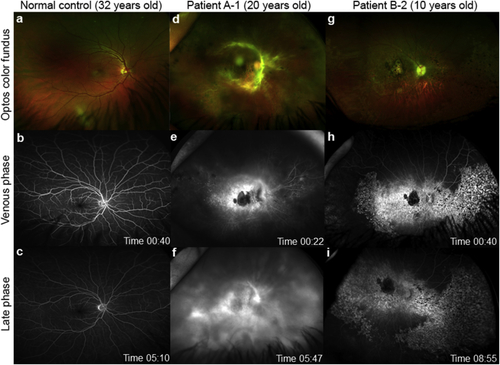
Fig. 3

Retinal vasculature in KCNJ13 patients.

Ultra-wide field fluorescein angiography (FFA) using Optos (Dunfermline, Scotland) of the right eye from two unrelated patients with a missense mutation (p.Thr153Ile) in KCNJ13 compared to normal control male age 32 years (a–c). Panel Patient A-1, 20 years old, (d) color fundus images showing vasoproliferative response around the arcades, widespread pan-retinal photocoagulation scars overlying the pigmentary retinopathy; (e) shows leakage (first seen in arterial phase, data not shown) in the venous phase with extensive leakage in the late phase (f). Panel Patient B-2, 10 years old, (g) color fundus images showing patchy areas of nummular pigmentary retinopathy; (h) FFA shows hyperfluorescence in areas of retinopathy with an area of hypofluorescence at the macula which coincides with delineated area of chorioretinal atrophy (choroidal vessels can be seen), but no evidence of leakage at any phase (i).

Expression Data

Expression Detail
Antibody Labeling
Phenotype Data

Phenotype Detail
Acknowledgments
This image is the copyrighted work of the attributed author or publisher, and ZFIN has permission only to display this image to its users. Additional permissions should be obtained from the applicable author or publisher of the image.

Reprinted from Experimental Eye Research, 189, Toms, M., Dubis, A.M., Lim, W.S., Webster, A.R., Gorin, M.B., Moosajee, M., Missense variants in the conserved transmembrane M2 protein domain of KCNJ13 associated with retinovascular changes in humans and zebrafish, 107852, Copyright (2019) with permission from Elsevier. Full text @ Exp. Eye. Res.