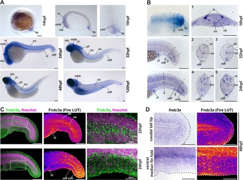
Localization of fndc3a RNA and protein during embryonic zebrafish development. (A,B) Expression of fndc3a mRNA was detected in the tail bud and the median fin fold from 14 hpf onwards. fndc3a is rather broadly expressed during embryogenesis, but was highly expressed in caudal and pectoral fins, somites, notochord cells and distinct brain regions. (C,D) Detection of Fndc3a protein via immunofluorescence indicated similar regional localization as fndc3a mRNA in 22–48 hpf embryos. Furthermore, it showed intracellular accumulation of Fndc3a at notochord cells, at somite boundaries and epidermal cells at this stage. In-situ stained embryos shown in (A,B) differ in proteinase K incubation and NBT/BCIP staining times to visualize weak fndc3a expression in different tissues and stages. Dashed lines in (B) indicate planes of the corresponding numbered sections 1–5, in (C) notochord boundary and in (D) fin fold border. Fire LUT in (C,D) shows pseudo-colored images of Fndc3a immunofluorescence and marks regions of high and low intensities (highest to lowest signal: yellow, red, blue, black). cnh: chordo neural hinge; cl: cloaca; le: lateral edge; mc: mesencephalon; mff: median fin fold; mhb: midbrain hindbrain boundary (marked with chevron); nk: neural keel; no: notochord; nt: neural tube; pf: pectoral fin; sb: somite boundary; so: somites; tb: tail bud; rc: rhombencephalon. Scale bars: 100 µm, except higher magnification in (C): 20 µm.
|
