|
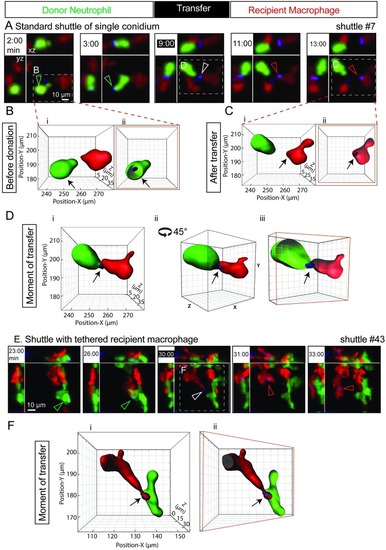
Shuttling of individual <italic>T</italic>. <italic>marneffei</italic> conidia from neutrophil to macrophage.(A) A representative standard shuttle of a calcofluor-stained conidium (blue) from a Tg(mpx:EGFP) neutrophil (green) to a Tg(mpeg1:Gal4FF)×(UAS-E1b:Eco.NfsB-mCherry) macrophage (red), corresponding to the example in S1A Movie. Panels include isometric orthogonal yz and xz views corresponding to the xy maximal intensity projection and indicate the time in min from start of movie. The time point colored white-on-black is the moment of transfer. Colored arrowheads indicate the conidium within donor neutrophil (green), at the point of intercellular transfer (white) and in the recipient macrophage (red). (B–D) Volume-rendered views of the standard shuttle in (A), detailed before (B), at the moment of transfer (C), and afterwards (D), demonstrating the intracellular location of the shuttled spore in donor neutrophil and recipient macrophage, the focal intercellular contact at the moment of transfer. Cii is the image in Ci rotated 45° around a central vertical axis in the direction shown. Images Bii, Ciii, and Dii are sectioned volume-rendered views; a sectioned plane is represented by a red box. (E–F) Shuttle demonstrating tethering of the departing recipient macrophage following a shuttle. Panel E presentation organized as in panel A. The tethered moment of transfer is detailed by volume-rendering in F, presented as in panels B–D. Scales as shown. Stills in A and E correspond to S1A and S1B Movie, respectively. Eco.Nfsb, Escherichia coli nitroreductase; EGFP, enhanced green fluorescent protein; Gal4FF, engineered form of Saccharomyces cerevisiae Gal4 transcriptional activator; mpeg1, macrophage-expressed gene 1; mpx, myeloid-specific peroxidase; Tg, transgenic; UAS-E1b, upstream activating sequence fused to minimal adenovirus E1b promoter.
|