|
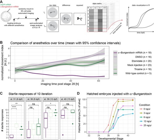
Anaesthesia of medaka is most efficient with α-Bungarotoxin mRNA injection.(A) Embryos were injected with eGFP mRNA or eGFP mRNA along with α-Bungarotoxin mRNA (1-cell stage modified from [17]). Injected embryos and uninjected embryos were dechorionated. Uninjected embryos were treated with Tricaine, Etomidate (experiments), DMSO or 1x ERM (controls). Embryos were randomly loaded in a 96-well plate and imaged automatically. Plates were adjusted by starting stage of imaging and fused into one dataset. Consecutively, image analysis was performed semi-automatically in FIJI and R. Therefore, the difference between each timepoint n and its following timepoint n+1 was calculated and squared. These values were imported into R and plotted along with their biological replicates. (B) Movement index of injected/treated embryos over time. Data was analysed as described in A. Strikingly, only α-Bungarotoxin is completely anesthetizing the embryos leading to nearly no detectable difference between the different timepoints (time resolution: 20 min, n as fish per condition is indicated in the legend). (C) α-Bungarotoxin-treated embryos are catching up in startle response ability. Fish were startled 10 times each and the number of responses was noted. Same legend as in Fig 4B applies. (α-Bungarotoxin: n = 12 fish, mock injected: n = 5 fish, wild-type control: n = 8 fish, asterisks indicate P-values: **** P < = 0.0001, *** P < = 0.001, ** P < = 0.01, * P < = 0.05, ns P > 0.05). (D) Different dilutions of α-Bungarotoxin were tested in order to adjust the concentration so fish would wake up after microscopy. Embryos were injected with 0, 3, 6, 12 or 25 ng/μl α-Bungarotoxin mRNA along with eGFP mRNA as an injection control. The number of hatched embryos per condition was scored (0 ng/μl: n = 33 fish, 3 ng/μl: n = 34 fish, 6 ng/μl: n = 16 fish, 12 ng/μl: n = 24 fish, 25 ng/μl: n = 22 fish).
|