|
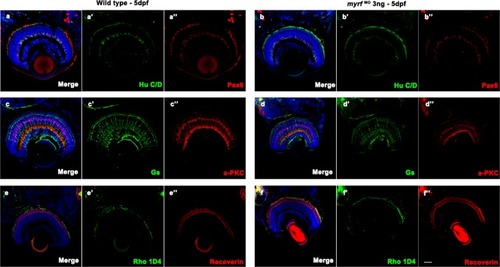
Immunostaining images of labels for different retinal cells in zebrafish at 5 dpf. Frozen sections from wild-type larvae (a–a″, c–c″, and e–e″) and myrf MO-injected larvae (b–b″, d–d″, and f–f″). Zebrafish larvae were labeled with Hu C/D (ganglion cell and amacrine cell marker, green, a′ and b′), Pax 6 (ganglion and amacrine precursor cell marker, red, a′′ and b′′), Gs (Müller cell marker, green, c′ and d′), PKCα (bipolar cell marker, red, c′′ and d′′), Rho 1D4 (long double cone outer segment marker, green, e′ and f′), recoverin (cone bipolar cell marker, red, e″ and f″), and DAPI (nuclei, blue). No differences were detected in retinal development between wild-type larvae and myrf morphants. Scale bars represent 100 μm
|