
Fig. 2
- ID
- ZDB-FIG-190826-6
- Publication
- Kague et al., 2019 - Scleraxis genes are required for normal musculoskeletal development and for rib growth and mineralization in zebrafish
- Other Figures
- All Figure Page
- Back to All Figure Page
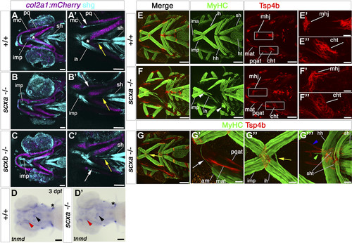
Cranial tendons, ligaments, and muscles of scxa−/− mutants are abnormal and disorganized. A–C) SHG imaging shows both collagen arrangement in tendons and ligaments and myosin heads in muscle (light blue) combined with confocal imaging of the transgene TgBAC(col2a1a:mCherry)hu5910 (cartilage, purple) of 7 dpf scxa−/−, scxb−/−, and wild-type (+/+) larvae. Shown are representative maximum projection images of the stacks (A–C) and single scans at comparable Z positions (A′–C′). Decreased SHG signal was observed in the ligaments connecting the Meckel’s and palatoquadrate cartilages of scxa and scxb mutants (white arrows), and in the sternohyoideus tendon (yellow arrows) connecting the sternohyoideus muscle and the basihyal cartilage (asterisk) of scxa mutants. D) In situ hybridization for tnmd for 3 dpf scxa−/− and their siblings (+/+) showing reduced tnmd mRNA levels in scxa mutant, such as the sternohyoideus tendon (black arrowhead) and the ligaments connecting the Meckel’s and palatoquadrate cartilages (red arrowhead). Expression at the base of the cleithrum is maintained (asterisk). E–G) Confocal stacks of 4 dpf embryos from a scxa+/− incross, immunostained for MyHC (A4.1025, green) and Tsp4b (red). Specific defects in tendon structure and directionality in scxa mutants (white rectangles) (E) are shown in higher magnification panels for mandibulohyoid junction (E′, F′) and ceratohyal tendon (E″, F″). Scxa mutants show varied array of muscle defects, such as abnormal overextension or crossing the midline of muscle fibers (F, G compared with E) and detached fibers in interhyoides and hyohyoides (arrowheads, F). The magnified area (G′–G″′) shows ectopic fiber in the adductor mandibularis (white arrow, G′), ectopic misguided fiber from the interhyoides is crossing the midline and growing toward the interhyoides across the midline (yellow arrow, G″), and ectopic extention of sternohyoides tendon (blue arrowhead) is attracting overextended fibers (green arrowhead). Am, adductor mandibularis; cht, ceratohyal tendon; hh, hyohyoides; ih, interhyoides; ima, intermandibularis anterior; imp, intermandibularis posterior; mat, Meckel’s adductor tendon; mc, Meckel’s cartilage; mhj, mandibulohyoid junction; pq, palatoquadrate cartilage; pqat, palatoquadrate adductor tendon; sh, sternohyoideus; sht, sternohyoides tendon. All images in ventral view, anterior to left. Scale bars, 100 µm, except F′–F″ and G′–G″′, 20 µm. |