|
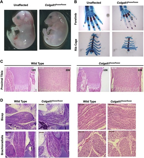
Loss of function of Colgalt1 leads to defects in musculoskeletal structure. (A) Colgalt1fosse/fosse embryos are smaller, rounded and swollen compared to unaffected littermates at E18.5. (B) Skeletal preparations of Colgalt1fosse/fosse embryos at E18.5 clearly demonstrate a reduction in the size of the bones in the wrist and the rib cage. (C) Hematoxylin and Eosin (H&E)-stained sections through the proximal tibiae indicate that there is no gross disorganization in the growth plates of this skeletal element at E18.5. (D) H&E-stained sagittal sections through the forelimbs show disorganized, ragged and shorter muscle fibers in the bicep and brachioradialis compared to wild type. Right hand panels show magnified views of sections in left hand panels. Scale bars: 200 μm (C, 10×; D, left) and 100 μm (C 20×; D, right).
|