|
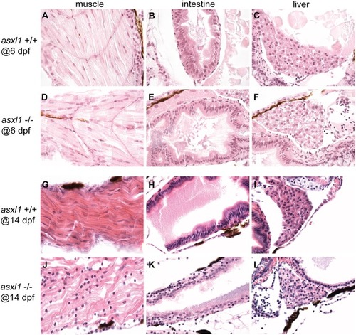
Asxl1 loss affects organ development. Histopathological analysis of the muscle, intestine and liver development with hematoxylin and eosin staining in 5 asxl1+/+ and 5 asxl1−/− mutants at 6 dpf and 14 dpf. Muscle (A,D) and intestine (B,E) development had no difference between asxl1+/+ and asxl1−/− embryos at 6 dpf. Liver parenchyma at 6 dpf appeared abnormal with vacuolated cells in asxl1−/− embryos (F) compared with asxl1+/+ embryos (C). At 14 dpf, muscular atrophy was shown in asxl1−/− embryos (J) compared with normal striated muscle in asxl1+/+ embryos (G). Intestine in asxl1−/− embryos was abnormal with villus blunting (K) compared with normal nuclei and microvilli in the asxl1+/+ embryos (H). The asxl1−/− embryos exhibited progressive liver architectural distortion (L) compared to asxl1+/+ embryos (I).
|