
FIGURE
Figure 1
- ID
- ZDB-FIG-190723-425
- Publication
- Li et al., 2019 - Methylglyoxal-Induced Retinal Angiogenesis in Zebrafish Embryo: A Potential Animal Model of Neovascular Retinopathy
- Other Figures
- All Figure Page
- Back to All Figure Page
Figure 1
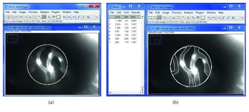
Quantification of the vascular area of retina at 4 dpf using Fiji-ImageJ software. An example of a retinal vascular image: use Fiji-ImageJ “area measurement” function under “Analyze” to measure the relative vascular vessel area over the retinal area. (a) Total retinal area by drawing a uniform circle of 120 pixel diameter = 11304 pixel area. (b) Draw and measure areas outside the vascular vessels = 2393 + 1256 + 638 + 1640 + 259 + 366 = 6552. Then, calculate % vascular vessel area of the retina = (11304 − 6552)/11304 × 100% = 42.0%. |
Expression Data
Expression Detail
Antibody Labeling
Phenotype Data
Phenotype Detail
Acknowledgments
This image is the copyrighted work of the attributed author or publisher, and
ZFIN has permission only to display this image to its users.
Additional permissions should be obtained from the applicable author or publisher of the image.
Full text @ J Ophthalmol