
Fig. 3
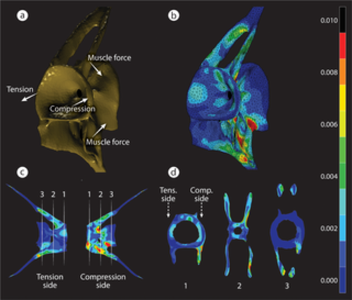
FEA of strain distribution in a loaded medaka vertebra. (a) A simplified loading graphic of the forces applied to the model. Lateral bending of the vertebral column during oscillatory swimming creates forces on the flat articular surfaces of adjacent centra, F Comp. on one side and F tens. on the opposite side. F Muscs acting on fish vertebrae are rather difficult to model because of the high complexity of the musculature as shown in S1 Fig and are represented here in a simplified manner. (b) 3D representation of von Mises strains in the vertebra. (c) Strain distribution in 2 contralateral views of a loaded vertebra. Locations of several transverse sections are marked by white dashed lines and are shown in (d). (d) Strain distribution in the 3 numbered 2D transverse sections shown in (c). Peak strains occur primarily near the external surfaces of the VB. Strain magnitudes (no units) for all images are shown in the color bar on the right. F Comp., compression force; FEA, finite element analysis; F Musc, muscle force; F Tens., tension force; VB, vertebral bone. |