
Fig. 3
- ID
- ZDB-FIG-190604-10
- Publication
- Lissouba et al., 2018 - Transcriptomic Analysis of Zebrafish TDP-43 Transgenic Lines
- Other Figures
- All Figure Page
- Back to All Figure Page
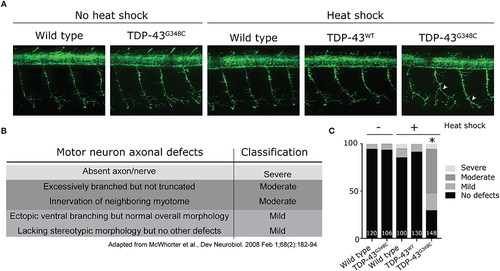
Abnormal development of motor neuron axons in zebrafish expressing TDP-43G348C. (A) Immunohistochemistry against znp-1 in 2 dpf whole-mount embryos. The 3-to-4 somites spanning the anus are shown with the axons of the motor neuron expanding down from the spinal cord. White arrow heads indicate abnormal premature branching of the main axonal branch and absence of secondary branching in transgenic embryos expressing TDP-43G348C, compared to embryos expressing TDP-43WT or not expressing any transgene. (B) Classification criteria used for the quantification of defects. (C) Quantification of axonal defects based on the classification found in (B). The total number of axons analyzed is indicated; The three axons spanning the anus were analyzed in at least 30 embryos over the course of three experiments. Chi-square analysis was performed between the different conditions, *p-value < 0.05. |
| Fish: | |
|---|---|
| Condition: | |
| Observed In: | |
| Stage: | Long-pec |