
FIGURE
Fig. 1
- ID
- ZDB-FIG-190306-2
- Publication
- Zhao et al., 2018 - Nux Vomica Exposure Triggered Liver Injury and Metabolic Disturbance in Zebrafish Larvae
- Other Figures
- All Figure Page
- Back to All Figure Page
Fig. 1
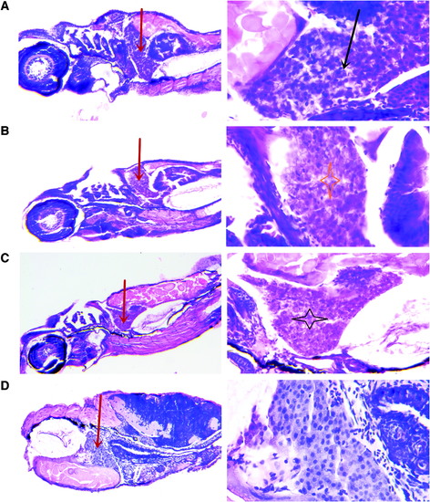
Histopathology of zebrafish liver after acute exposure to nux vomica for 24 h. Photomicrographs of liver sections (4–5 μm) stained with hematoxylin and eosin. (A) Livers from zebrafish exposed to 30 μg/mL. A number of hepatocytes were swollen (black arrow). (B) The hepatocyte borders became obscure, and cell vacuolar degeneration was noted (asterisk) in the 50 μg/mL group. (C) Destruction of cellular structures and hepatocyte denaturation were observed (asterisk). (D) Livers from the control group. The red arrow is the liver in the different group. Magnification 100 × and 400 × . |
Expression Data
Expression Detail
Antibody Labeling
Phenotype Data
Phenotype Detail
Acknowledgments
This image is the copyrighted work of the attributed author or publisher, and
ZFIN has permission only to display this image to its users.
Additional permissions should be obtained from the applicable author or publisher of the image.
Full text @ Zebrafish