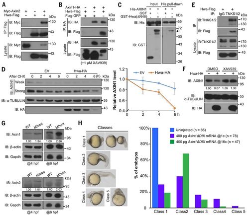
Hwa binds to and promotes the degradation of Axin. (A and B) Hwa-Flag physically interacts with Myc-Axin2 (A) and Axin1-HA (B) in HEK293T cells. Zebrafish Hwa and mouse Axin1 and Axin2 were used. XAV939 is a tankyrase inhibitor. Note that Axin1 and Axin2 levels are reduced when zebrafish Hwa is expressed. (C) Direct binding of Hwa with AXIN1. His-AXIN1 (N351) protein of human AXIN1 origin was incubated with GST-Hwa(ΔΝ46) protein of zebrafish origin expressed in E. coli. GST-Hwa (indicated by an arrow) is detected by Western blotting in the His-AXIN1 precipitates. (D) Hwa-HA promotes degradation of endogenous AXIN1 in HEK293T cells. Left: Cells transfected with empty vector (EV) or Hwa-HA were treated with cycloheximide (CHX; 100 μg/ml) and harvested for immunoblotting at indicated time points. Right: Dynamic change of AXIN1 protein level (normalized to α-TUBULIN), shown as mean ± SD. (E) Hwa-Flag binds to endogenous TNKS1/2 in HEK293T cells. (F) Hwa-promoted degradation of endogenous AXIN1 in HEK293T cells is prevented by inhibition of tankyrases. Six hours after transfection, cells were treated with DMSO or 1 μM XAV939 for 18 hours. Relative AXIN1 levels (normalized to α-TUBULIN) are indicated. (G) Immunoblotting reveals an increase of endogenous Axin1 and Axin2 proteins in Mhwatsu01sm mutants at 4 and 6 hpf. Relative levels are indicated. (H) Axin1ΔDIX overexpression rescues the body axes of Mhwatsu01sm mutants. Axin1ΔDIX mRNA was injected into one-cell stage mutants or into one blastomere of 16-cell stage mutants. Embryos at 24 hpf were categorized into five classes (left) and the ratio of embryos in each class is shown (right).
|
