
Fig. S10
- ID
- ZDB-FIG-181207-37
- Publication
- Tessadori et al., 2018 - Effective CRISPR/Cas9-based nucleotide editing in zebrafish to model human genetic cardiovascular disorders
- Other Figures
- All Figure Page
- Back to All Figure Page
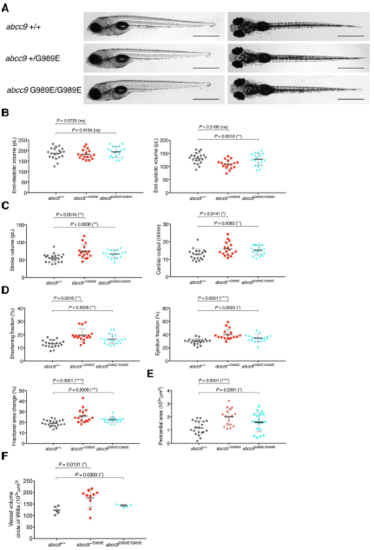
abcc9[G989E] embryos show CS-related anomalous heart function and pericardial edema at 5 dpf. (A) Comparison of wild-type and abcc9[G989E] mutants at 5 dpf seen from a left lateral (left) and dorsal view (right). (B-D) Quantification of cardiac function using sequential still frames from high-speed imaging of the embryonic cardiac cycle at 5 dpf. Ventricular volume (B), cardiac output (C) and contractile function (D) were calculated. (E) Quantification of pericardial edema by measuring pericardial area using striking morphological landmarks. In order to correct for possible enlarged ventricular size in mutants, ventricular area was subtracted. (F) Quantification of vascular dilations in a Tg(kdrl:GFP) background. 3D reconstruction of vascular structure in Imaris was used to calculate vessel volume. For all graphs, significance was determined by two-tailed unpaired Student's t test or Mann–Whitney two-tailed U test: * p≤0.05; ** p≤0.01; *** p≤0.001; **** p≤0.0001. The black horizontal bar indicates the mean value for each condition. Sample size, abcc9+/+, n=20; abcc9+/G989E, n=17; abcc9G989E/G989E, n=19 in B-E; abcc9+/+, n=5; abcc9+/G989E, n=10; abcc9G989E/G989E, n=5 in F. Scale bar, 1 mm in A. All embryos analyzed originated from group matings of adult zebrafish. |
| Fish: | |
|---|---|
| Observed In: | |
| Stage: | Day 5 |