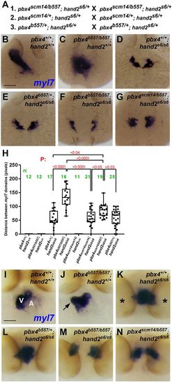
The pbx4 p.A131V variant enhances myocardial morphogenesis defects caused by loss of hand2. (A) Genetic crosses of zebrafish strains used to obtain embryos for the analyses of pbx4;hand2 mutant embryos. All adult breeder fish used in crosses 1-3 were ‘siblings’, derived from the same clutch from a group cross. (B-G) Myocardial marker myl7 expression at 24 hpf. Dorsal views; anterior is up. Animal numbers for phenotypic classes are provided in Table 2. (B) The heart tube appears normal in pbx4+/+;hand2+/+ embryos. (C) pbx4b557/b557;hand2+/+ embryos have a medial heart cone myl7 domain. (D) pbx4+/+;hand2s6/s6 embryos show a crescent-shaped myocardial fusion defect of the myl7 domains. (E) A similar phenotype is observed in pbx4b557/+;hand2s6/s6. (F,G) The myl7 fusion defect is more severe in pbx4b557/b557;hand2s6/s6 (F) and pbx4scm14/b557;hand2s6/s6 (G). (H) Quantitation of fusion defect of myl7 domains in different genetic combinations. myl7 distance measurements were made blind to embryo genotypes. The averages for the myl7 distances among the different genotypes were compared using one-way ANOVA, and P-values were corrected for multiple comparisons using Tukey's test. The boxes extend from the 25th to 75th percentiles, the whiskers are at the minimum and maximum, and the bar within the box represents the median. (I-N) Myocardial marker myl7 expression at 60 hpf. In I-J, ventral views; anterior is up. In K-N, anterior views; dorsal is up. Animal numbers for phenotypic classes are provided in Table 3. (I) The heart appears normal in pbx4+/+;hand2+/+ embryos. V, ventricle; A, atrium. (J) pbx4b557/b557;hand2+/+ embryos have dysmorphic hearts with bulges of the ventricular myocardium (arrow). (K) pbx4+/+;hand2s6/s6 embryos show an abnormally shaped, medial myocardium positioned more caudally between the eyes (asterisks). (L) A similar phenotype is observed in pbx4b557/+;hand2s6/s6. (M,N) More severe myl7 bilateral domain phenotypes are observed in pbx4b557/b557;hand2s6/s6 (M) and pbx4scm14/b557;hand2s6/s6 (N) embryos. Scale bars: 50 μm.
|
