
Fig. 3
- ID
- ZDB-FIG-181101-3
- Publication
- Willoughby et al., 2012 - Generation of a genetically encoded marker of rod photoreceptor outer segment growth and renewal
- Other Figures
- All Figure Page
- Back to All Figure Page
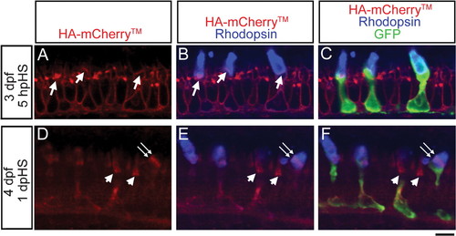
Expression of HA-mCherryTM in photoreceptors after heat-shock at 3 dpf. (A–C) At 5 hpHS, a single confocal z-section of a Tg(hsp70:HA-mCherryTM) photoreceptor layer labeled with anti-HA antibody (red), anti-Rhodopsin antibody (blue) and GFP-expressing rods (green) shows membrane expression of HA-mCherryTM in rods and neighboring cones, and a stripe of HA-mCherryTM at the base of rod outer segments (arrows, A, B). (D–F) At 1 dpHS, a single confocal z-section of a Tg(hsp70:HA-mCherryTM) photoreceptor layer labeled with anti-HA antibody (red), anti-Rhodopsin antibody (blue) and GFP-expressing rods (green) shows that membranous HA-mCherryTM labeling in the cell body and inner segment has largely disappeared and a stripe of HA-mCherryTM in a rod outer segment has been displaced distally (double arrows, D–F). Continuous HA-mCherryTM labeling largely fills cone outer segments (arrowheads, D–F). Scale bar: A–F, 5 µm. |