
Fig. 7
- ID
- ZDB-FIG-181011-14
- Publication
- Li et al., 2018 - Mutation in the intracellular chloride channel CLCC1 associated with autosomal recessive retinitis pigmentosa
- Other Figures
- All Figure Page
- Back to All Figure Page
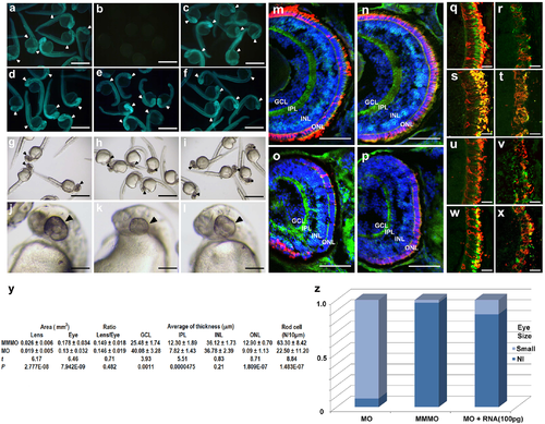
Zebrafish eye development disturbed by knockdown of clcc1 expression. Validation: Injection of the 5’-modified EGFP (a) or the unmodified EGFP (d) gave a fluorescent signal (arrowheads). Co-injection of the clcc1-MO eliminated the fluorescent signal from morpholino-sensitive 5’-modified EGFP mRNA (b) but not unmodified EGFP (e). Co-injection of the MM-MO had no effect (c, f). Eye Size: Injection of the clcc1-MO (h, k) significantly reduced eye size (black arrows) compared to MM-MO (i, l) and buffer-injected (g, j) embryos. a-f: 24 hpf, (g-l): 36 hpf. Retinal frozen sections: from 4 dpf MM-MO- (m, n) and clcc1-MO-injected (o, p) embryos stained for PKCß1 (bipolar cells, green), Zpr-1 (cone receptors, red, n and p), 1D1 (rod receptors, red, m and o), and DAPI (nuclei, blue). clcc1-MO-injected embryos show decreased thickness of ONL and IPL layers. MM-MO-injected (q, s, u, w) and clcc1-MO-injected (r, t, v, x) embryos were stained with anti-blue opsin (q, r, green), anti-green opsin (s, t, green), anti-red opsin (u, v, green), or anti-UV opsin (w, x, green), and 4D2 (all, Rhodopsin, rods, red). All photoreceptors in clcc1-MO-injected embryos show reduced staining and damaged photoreceptor cell structure, with the greatest decreases in blue and green opsin cones. m-x: 4 dpf. Scale Bar: m-p: 50 μm, q-x: 10 μm. Comparison of eye size and retinal layers: (y). GCL = ganglion cell layer, IPL = inner plexiform layer, INL = inner nuclear layer, and ONL = outer nuclear layer. Proportions of embryos with eye size phenotype: (z) with clcc1-MO injection and rescue by coinjected clcc1 WT mRNA. Lens and eye areas are given in mm2, and retinal thicknesses are given in μm. Forty clcc1-MO-treated embryos and 41 MM-MO-treated embryos were analyzed. |
| Antibodies: | |
|---|---|
| Fish: | |
| Knockdown Reagent: | |
| Anatomical Terms: | |
| Stage: | Day 4 |
| Fish: | |
|---|---|
| Knockdown Reagent: | |
| Observed In: | |
| Stage Range: | Prim-5 to Day 4 |