
Fig. S1
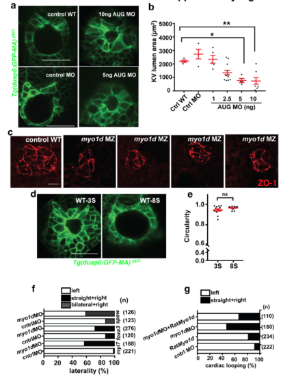
myo1d is necessary for lumen morphogenesis of Kupffer’s vesicle a. Representative images showing KV lumen surface area with increasing doses of myo1d AUG MO injected into Tg(dusp6:GFP-MA) embryos. b. Graph showing KV lumen formation was significantly decreased with increasing doses of myo1d AUG MO injected into embryos. c. Loss of Myo1d leads to dysmorphic lumen formation. ZO1 staining marks apical surface in myo1d MZ embryos showing smaller or dysmorphic KV shape when compared to controls. scale bar-20μm. d. Representative images of Tg(dusp6:GFP-MA) embryos showing KV formation at 3S and 8S stages used to calculate circularity. e. Graph showing circularity of KVs from 3S and 8S embryos. f. Laterality, expressed as percentage, from MO injected embryos. Embryos were fixed and probed using RNA in situ probes for myl7, foxa3 and spaw, and scored for defective looping pattern. Knockdown of Myo1d increased laterality defects. g. Cardiac looping, expressed as percentage from MO injections and rescue with rat Myo1d mRNA. Injected embryos fixed and probed for myl7 expression as a marker for cardiac looping. Knockdown of myo1d resulted in looping defects that was reduced with co-injection of rat Myo1d mRNA. |