
Fig. 4
- ID
- ZDB-FIG-180913-67
- Publication
- de Vos et al., 2018 - Functional analysis of a hypomorphic allele shows that MMP14 catalytic activity is the prime determinant of the Winchester syndrome phenotype
- Other Figures
- All Figure Page
- Back to All Figure Page
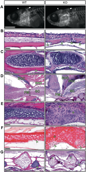
The mmp14a/b KO zebrafish have abnormal enchondral and membranous ossifying skull bones and Weberian vertebrae. (A) Fluorescence microscopy images of 30 dpf WT and mmp14a/b KO juveniles, whole mount stained for calcified bone with alizarin red; lateral view, anterior to the left. At 30 dpf, the frontal bones (arrowhead) and the supraoccipital bone (SOC, arrow) of mmp14a/b KO fish are shaped differently compared with age and size-matched (10.2 mm standard length) WT fish. (B–E) H&E stained mid-sagittal sections of 90 dpf fish; anterior to the left except for the mmp14a/b KO section shown in (E), which is rotated (anterior at the bottom) for clearer comparison with the corresponding WT section. At 90 dpf, the frontal bones (B) of mmp14a/b KO fish are irregularly thickened and contain cell clusters (arrowheads). The dentary bone (C) and SOC (E) of mmp14a/b KO fish contain a relative large amount of disorganized cartilage and small amounts of bone matrix (arrowheads). The SOC additionally shows cell-free areas that have lost basophilia, indicating lack of proteoglycans (E, asterisks). The SOC and second supraneural (SN2) form a sharp angle and are ventrally extended in mmp14a/b KO fish, impinging the spinal cord (SC) at the foramen magnum (FM, compare diameter indicated by black line in D). (F) Sagittal picrosirius red stained sections of the same fish as shown in (E) (same orientation as in (E) reveal the SOC of mmp14a/b KO fish lacks a collagen-rich peripheral bone matrix, but instead contains large cell-rich areas (arrowheads) as compared with WT fish. In contrast, cell-free regions in the cartilage core of mmp14a/b KO fish are relatively intensely stained. (G) H&E stained mid-sagittal sections (anterior to the left) demonstrating Weberian vertebral bodies of 90 dpf mmp14a/b KO fish are irregularly shaped and contain cell clusters (arrows), while the intervertebral cartilage is absent (arrowhead) compared with WT fish. Scale bar in (A) equals 1 mm, scale bars in (B), (C), (E) and (F) equal 20 µm, scale bar in (D) equals 200 µm, scale bar in (G) equals 100 µm. |
| Fish: | |
|---|---|
| Observed In: | |
| Stage Range: | Days 30-44 to Adult |