
Fig. 3
- ID
- ZDB-FIG-180912-3
- Publication
- Gurung et al., 2018 - Distinct roles for the cell adhesion molecule Contactin2 in the development and function of neural circuits in zebrafish
- Other Figures
- All Figure Page
- Back to All Figure Page
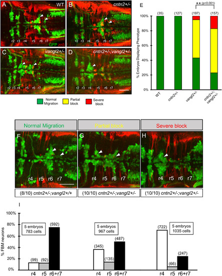
cntn2 interacts genetically with vangl2 but not with lamc1. Panels A–D and F–H show dorsal views of the hindbrain with anterior to the left. Tg(isl1:gfp) embryos were fixed at 48 hpf, and processed for immunohistochemistry with zn5 antibody (red) to label hindbrain commissural neurons and axons at rhombomere boundaries, and anti-GFP antibody (green) to label FBM neurons (arrowheads). (A) FBM neurons migrate normally in a control embryo. (B) FBM neurons migrate normally in a cntn2 heterozygous (cntn2+/−) embryo. (C) FBM neurons migrate poorly in a vangl2 heterozygous (vangl2+/−) embryo, with neurons located along the entire migratory pathway from r4 to r6. (D) FBM neurons fail to migrate out of r4 in a cntn2; vangl2 double heterozygote (cntn2+/−; vangl2+/−). Scale bar in D, 50 μm for A–D. (E) Quantification of genetic interaction data. Number in parenthesis denotes number of embryos. **Chi-square test at p < 0.001; NS: not significant. Data are from 2 to 4 experiments. (F–H) Offsprings of vangl2+/− heterozygous and cntn2−/− homozygous mutants exhibit normal, partial block, and severe block phenotypes for FBM neuron migration. Embryos exhibiting partial block (10/10) and severe block (10/10) were all identified as cntn2; vangl2 double heterozygote (cntn2+/−; vangl2+/−) and a majority of embryos (8/10) exhibiting normal migration were identified as cntn2+/−; vangl2+/+ by genotyping. (I) Quantification of non-migrated FBM neurons in r4, partially migrated FBM neurons in r5 and fully migrated FBM neurons in r6, and r7. Number in parenthesis denotes number of cells. Scale bar in F, 50 μm for F–H. |
| Gene: | |
|---|---|
| Antibody: | |
| Fish: | |
| Anatomical Terms: | |
| Stage: | Long-pec |
| Fish: | |
|---|---|
| Observed In: | |
| Stage: | Long-pec |
Reprinted from Mechanisms of Development, 152, Gurung, S., Asante, E., Hummel, D., Williams, A., Feldman-Schultz, O., Halloran, M.C., Sittaramane, V., Chandrasekhar, A., Distinct roles for the cell adhesion molecule Contactin2 in the development and function of neural circuits in zebrafish, 1-12, Copyright (2018) with permission from Elsevier. Full text @ Mech. Dev.