
Fig. 5
- ID
- ZDB-FIG-180912-18
- Publication
- Kinoshita et al., 2018 - Functional roles of the Ripply-mediated suppression of segmentation gene expression at the anterior presomitic mesoderm in zebrafish
- Other Figures
- All Figure Page
- Back to All Figure Page
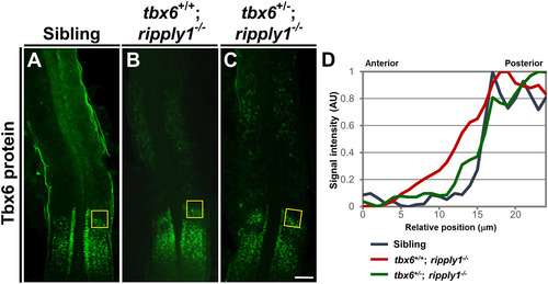
Immunostaining of Tbx6 protein in the PSM of sibling, tbx6+/+; ripply1−/−, and tbx6+/−; ripply1−/− embryos. Immunohistochemistry was carried out using an anti-Tbx6 antibody. Sibling (A), tbx6+/+; ripply1−/− (B), and tbx6+/−; ripply1−/− (C) embryos at the 15-somite stage were examined. The tbx6+/−; ripply1−/− embryos that clearly exhibited recovered somite boundaries were used for analysis. Deyolked embryos were flat-mounted on slide glasses. Dorsal views and the anterior is to the top. Scale bar is 100 μm. (D) Spatial qualifications of Tbx6 signal around the anterior border of Tbx6 domains in the PSM of sibling, tbx6+/+; ripply1−/−, and tbx6+/−; ripply1−/− embryos. In the boxed areas shown in A, B, and C, the intensity of Tbx6 signals along the anterior-posterior axis was measured by ImageJ software, and the relative intensity was shown. |
| Gene: | |
|---|---|
| Fish: | |
| Anatomical Terms: | |
| Stage: | 14-19 somites |
| Fish: | |
|---|---|
| Observed In: | |
| Stage: | 14-19 somites |
Reprinted from Mechanisms of Development, 152, Kinoshita, H., Ohgane, N., Fujino, Y., Yabe, T., Ovara, H., Yokota, D., Izuka, A., Kage, D., Yamasu, K., Takada, S., Kawamura, A., Functional roles of the Ripply-mediated suppression of segmentation gene expression at the anterior presomitic mesoderm in zebrafish, 21-31, Copyright (2018) with permission from Elsevier. Full text @ Mech. Dev.