
Fig. 6
- ID
- ZDB-FIG-180824-43
- Publication
- Karthik et al., 2018 - Synergistic interaction of sprouting and intussusceptive angiogenesis during zebrafish caudal vein plexus development
- Other Figures
- All Figure Page
- Back to All Figure Page
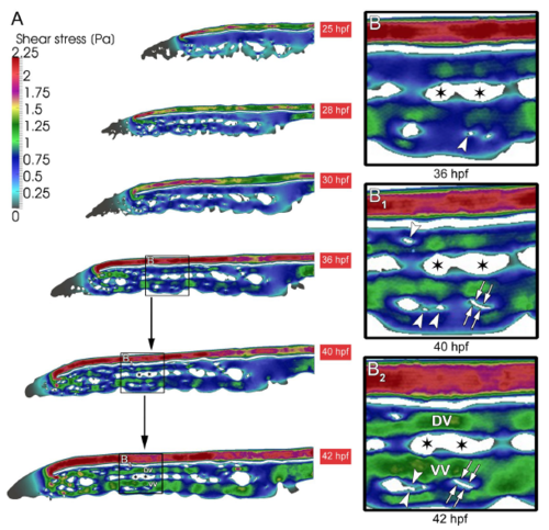
Shear stress distribution during CVP development. (A) The overall shear stress distribution calculated from blood flow videos obtained from in vivo microscopy between 25-42 hpf of developing CVP. (B,B 1 ) Arrowheads show the appearance of pillars in the regions of declining shear stress. (B 1 ,B 2 ) Subsequently the same shear stress profile is associated with the path of pillar fusion indicated in white double arrows leading to the splitting of the vessel. The ensuing splitting of the vessels between 36-42 hpf leading to the formation of dorsal (DV) and ventral vein (VV). The splitting path is indicated with asterisks. |