
Fig. 7
- ID
- ZDB-FIG-180522-11
- Publication
- Kimelman et al., 2017 - Regulation of posterior body and epidermal morphogenesis in Zebrafish by localized Yap1 and Wwtr1
- Other Figures
- All Figure Page
- Back to All Figure Page
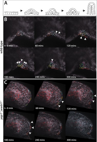
Dynamic movements of presumptive epidermal cells at the ventral fin fold. (A) Cartoon showing previous analysis of dorsal fin fold formation redrawn from Dane and Tucker, 1985. (B) Stills from live imaging of ventral fin fold formation in wild-type embryos taken from Video 2. (C) Stills from live imaging of ventral fin fold formation in yap1;wwtr1 double mutants taken from Video 3. Images are of the midline with anterior to the left. Arrowheads point to new cells arriving at the midline. Most of the cells in the wild-type arrive basally, although two cells outlined appear at the edge of the fin fold coming from the other side of the embryo. Note that in wild-type embryos most cells that are at the midline at t = 0 min, or come to the midline at later times, stay at the midline whereas in the yap1;wwtr1 double mutants cells are frequently transiently at the midline (quantified in Figure 8). Lateral views of the ventral-posterior part of the embryo, with dorsal up and ventral down. Wild-type movie is a representational movie of 21 movies examined and the mutant movie is representational of 11 movies. |
| Fish: | |
|---|---|
| Observed In: | |
| Stage Range: | 20-25 somites to 26+ somites |