
Fig. S5
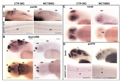
Expression of differentially expressed genes (p<0.01; FDR 5%) involved in zebrafish neural development. Comparison between control and MCT8 morphant zebrafish embryos at 48hpf. (A) WISH expression analysis of neural progenitor marker pax6a, this gene is regulated in a context dependent manner by MTHs during zebrafish embryogenesis. Lateral images of the hindbrain and spinal cord of analysed embryos are presented (B) WISH expression analysis of neural progenitor factor neurod6b. This gene is regulated by MTH in the mid- and hindbrain. Lateral (upper panel) and dorsal images (lower panel) of the brain of analysed embryos is presented. hb-hindbrain, (C) WISH expression analysis of Retinoic orphan receptor ab (rorab). Regulation by MTH occurs in the midbrain and eyes. Lateral (first panel) and dorsal images (second panel) of the brain of analysed embryos are presented. Red arrowheads indicate the optic tectum. (D) WISH analysis of expression of inhibitory neuron marker gad1b, showing that inhibitory neurons development is dependent of MTHs in zebrafish embryogenesis. Lateral and dorsal images of brain (first and second panels) of analysed embryos are presented and lateral images of spinal cord are shown (lower panel). Red arrowhead indicates the midbrain-hindbrain boundary (MHB). nt-notochord, md – midbrain, fb – forebrain, ey – eye. In all images scale bar represents 100μm |