
Fig. s4
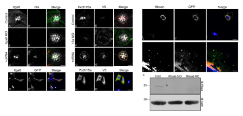
Qualification of the antibody preparations. A-D’: Guinea pig antibodies against an extracellular peptide sequence (REFESKPREVGRVYLY) of Itga8 were qualified in 3dpf MOs (A-C’) and HEK293 cells (D-D’). Confocal images of controls (AA’), itga8 MOs (B-B’) or itga8 MOs+cRNA coding for Itga8-His (C-C’), showing immunostaining for Itga8 and His6x. D-D’: Confocal images of HEK293 transiently electroporated with Itga8-GFP and immunostained with anti-Itga8 and anti-GFP. E-H’: Rabbit antibodies against a carboxyl-terminal peptide (KNSDRFGCSPDVKYDNKNDV) of Pcdh15a (Pcdh15a-CD1) were qualified in 3dpf larvae (E-G’) and HeLa cells (H-H’). Confocal images of controls (E-E’), pcdh15a MOs (F-F’) or pcdh15a MOs+cRNA coding for Pcdh15a-V5 (G-G’) showing immunostaining for Pcdh15a and V5 epitope. H-H’: Confocal images of HeLa cells transiently electroporated with Pcdh15a-V5 and immunostained with anti-Pcdh15a and anti-V5. I-J’: Mouse monoclonal antibodies against a peptide sequence common to both Rhoa proteins (VADIEVDSKQVELAC) were qualified in HeLa cells transiently electroporated with Rhoab-GFP. Confocal images showing immunostaining with anti-Rhoa and anti-GFP. J-J’: High magnification of the boxed area in I-I’, showing positive perinuclear and vesicular immunostaining. Similar results were obtained with Rhoad-GFP (data not shown). A-C’, E-G’: hair cells were counterstained with phalloidin (merged images). D-D’, H-H’, I-I’: DAPI was used to counterstain the nucleus. Asterisks denote untransfected cells used as control. Scale:A-C’, E-G’: 5.5µm; D-D’, H-H’: 19.5µm; I-I’: 17µm; J-J’: 2.5µm. K: Rhoa immunoblot of 3dpf controls, Rhoab MOs (5.7ng) and Rhoad MOs (8ng). Asterisk denotes specific band. Membrane was stripped and blotted for actin as a loading control. At least three independent experiments were performed with the morphants and at least two independent experiments with the cell cultures. Re-localization (and re-expression) of the Itga8 or Pcdh15a proteins in the MOs coinjected with the full length cRNA, was confirmed by co-labeling with the corresponding antibody and anti-tag antibodies (anti-His for Itga8 and anti-V5 for Pcdh15a), demonstrating the specificity of the knockdown and also, precluding the possibility of interference between the morpholino suspension and the cRNA as it has been suggested by Kok et al. (2015). |