
Fig. 5
- ID
- ZDB-FIG-180308-24
- Publication
- Chen et al., 2017 - Imaging early embryonic calcium activity with GCaMP6s transgenic zebrafish
- Other Figures
- All Figure Page
- Back to All Figure Page
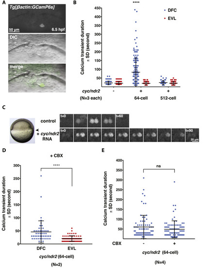
Excess Nodal signaling prolongs Ca2+ transient duration specifically in the DFCs. (A) Representative images of a single Ca2+ transient in a DFC at 6.5 hpf. (B) Quantification of Ca2+ transient duration in WT and Cyc/Ndr2-misexpressing embryos. Error bars represent standard deviation. ****, P≤0.0001. N=3 embryos in control and Cyc/Ndr2-misexpressing embryos, respectively. (C) Still images of Ca2+ transients in the DFCs of uninjected control and Cyc/Ndr2-misexpressing embryo in a time-lapse series. Arrowhead points to the DFC region that was imaged. (D) Quantification of DFC Ca2+ transient duration between DFCs and EVL cells in cyc/ndr2 RNA-injected embryos after CBX treatment. ****, P≤0.0001. N=2 embryos. (E) Quantification of DFC Ca2+ transient duration in Cyc/Ndr2-misexpressing embryos before and after CBX treatment. ns, not significant; N=4 embryos. |
Reprinted from Developmental Biology, 430(2), Chen, J., Xia, L., Bruchas, M.R., Solnica-Krezel, L., Imaging early embryonic calcium activity with GCaMP6s transgenic zebrafish, 385-396, Copyright (2017) with permission from Elsevier. Full text @ Dev. Biol.