
FIGURE
Fig. 3
- ID
- ZDB-FIG-180308-1
- Publication
- Shin et al., 2017 - PRAJA is overexpressed in glioblastoma and contributes to neural precursor development
- Other Figures
- All Figure Page
- Back to All Figure Page
Fig. 3
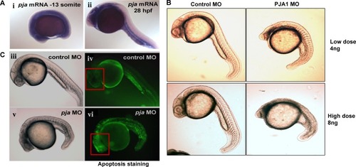
A. PJA1 mRNA expression was determined by whole-mount in situ hybridization in developing embryos. PJA1 mRNA is expressed in the yolk and ventral side of the embryo at the 13 somite stage (i) and in intestinal and vascular tissues at 28 hour post-fertilization (hpf) (ii). B. Developmental defects were observed in zebrafish embryos injected with 4ng or 8ng of antisense PJA1 morpholino oligonucleotides (MO) but not in embryos injected with same dose of control MO. C. Apoptosis was visualized by acridine orange staining in live embryos, where apoptotic cells are indicated by green fluorescence. Compared to that of the control MO injected embryos at 24 hpf. D. injection of PJA1 MOs severely disrupted embryogenesis (v) and resulted in high levels of apoptosis (vi). Red squares indicate the embryonic brain of zebrafish.
|
Expression Data
| Gene: | |
|---|---|
| Fish: | |
| Anatomical Terms: | |
| Stage Range: | 5-9 somites to Prim-5 |
Expression Detail
Antibody Labeling
Phenotype Data
| Fish: | |
|---|---|
| Knockdown Reagent: | |
| Observed In: | |
| Stage: | Prim-5 |
Phenotype Detail
Acknowledgments
This image is the copyrighted work of the attributed author or publisher, and
ZFIN has permission only to display this image to its users.
Additional permissions should be obtained from the applicable author or publisher of the image.
Full text @ Genes Cancer