
Fig. 2
- ID
- ZDB-FIG-171113-30
- Publication
- Powell et al., 2016 - Zebrafish Müller glia-derived progenitors are multipotent, exhibit proliferative biases and regenerate excess neurons
- Other Figures
- All Figure Page
- Back to All Figure Page
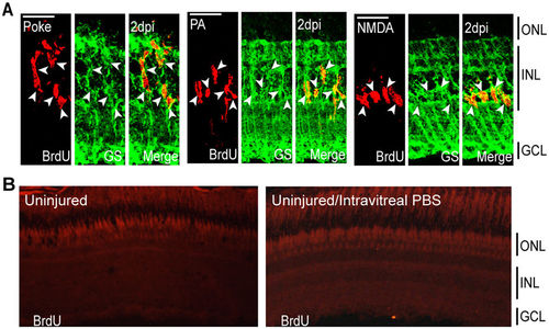
Injury models stimulate MG proliferation by 2 dpi in the INL. (A) Representative confocal images of retinal sections immunostained for BrdU and GS at 2 dpi following needle poke, PA, and NMDA injuries. Fish were given an injection of BrdU intraperitoneally 3 hours before harvest. Scale bar is equal to 50 μm. (B) PBS was injected into the vitreous of eyes whose retinas were uninjured. Fish were then given an injection of BrdU intraperitoneally 3 hours before harvest at 2 days post PBS injection. Shown are representative images of BrdU immunofluorescence in retinal sections. Similar results were obtained when BrdU was injected at 4 days post PBS injection. ONL, outer nuclear layer; INL, inner nuclear layer; GCL, ganglion cell layer; PA, photoablation; GS, glutamine synthetase; dpi, days post injury. |