
Fig. 3
- ID
- ZDB-FIG-170914-47
- Publication
- Tornini et al., 2017 - Live fate-mapping of joint-associated fibroblasts visualizes expansion of cell contributions during zebrafish fin regeneration
- Other Figures
- All Figure Page
- Back to All Figure Page
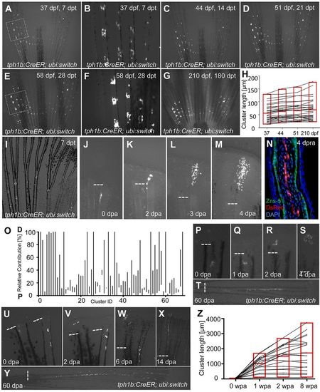
tph1b+ fibroblasts contribute to peri-joint regions in the absence of injury but broaden contributions during regeneration. (A-G) tph1b:CreER; ubi:switch fish were treated with 4 µM tamoxifen at 30 dpf, and labeled cells in caudal fins were tracked at 7 (A,B), 14 (C), 21 (D), 28 (E,F) and 180 (G) days post-tamoxifen treatment (dpt). The boxed areas in A and E are magnified in B and F, respectively, showing limited contributions of tph1b+ cells and progeny to local joint tissue throughout adulthood. (H) Plot of tph1b+ proximodistal (PD) cluster lengths (in µm) during normal growth measured at 37, 44, 51 and 210 dpf. tph1b:CreER; ubi:switch fish were treated as in A. n=31 clusters. Labeled cells remained closely associated to joints. Red box indicates minimum/maximum values, with the average value in the middle. (I) An uninjured adult tph1b:CreER; ubi:switch caudal fin, treated with 4 µM tamoxifen and imaged at 7 dpt. tph1b+ cells near joints are labeled. (J-M) Contribution of a labeled tph1b+ cell in tph1b:CreER; ubi:switch fish, shown at 0 (J), 2 (K), 3 (L) and 4 (M) dpa. Dashed line indicates amputation plane. (N) Longitudinal section of a tph1b:CreER; ubi:switch fin regenerate at 4 days post-re-amputation (dpra), stained for osteoblasts (ZNS-5, green), mCherry (DsRed, red) and nuclei (DAPI, blue). We saw no evidence for contributions by tph1b+ joint-associated cells to osteoblasts during regeneration. (O) Plot of final relative position along the PD axis in which tph1b+ cell progeny are distributed, normalized for regenerated ray length as a percentage (0, proximal; 100, distal). Fins were amputated near labeled tph1b+ cells and progeny were tracked through 60 dpa. n=68. P, proximal; D, distal. (P-T) Contribution of a tph1b+ labeled cell in tph1b:CreER; ubi:switch fish, shown at 0 (P), 1 (Q), 2 (R), 4 (S) and 60 (T) dpa. Joint-associated tph1b+ cells can contribute progeny to regenerated tissue well beyond joint areas. (U-Y) Contribution of a tph1b+ labeled cell in tph1b:CreER; ubi:switch fish (left ray), shown at 0 (U), 2 (V), 6 (W), 14 (X) and 60 (Y) dpa. Joint-associated tph1b+ cells can contribute progeny across the entire PD axis of the regenerate. (Z) Plot of tph1b+ progeny lengths (in µm) during fin regeneration. Adult tph1b:CreER; ubi:switch fish were treated as in I. At 7 dpt, fins were amputated distal to labeled cells, and progeny lengths were measured at 0, 1, 2 and 8 weeks post-amputation (wpa). n=20. Labeled cells significantly expanded their contributions. |
| Gene: | |
|---|---|
| Fish: | |
| Conditions: | |
| Anatomical Terms: | |
| Stage: | Adult |