
FIGURE
Fig. S6
- ID
- ZDB-FIG-170424-15
- Publication
- Schmitner et al., 2017 - ptf1a+ , ela3l- cells are developmentally maintained progenitors for exocrine regeneration following extreme loss of acinar cells in zebrafish larvae.
- Other Figures
- All Figure Page
- Back to All Figure Page
Fig. S6
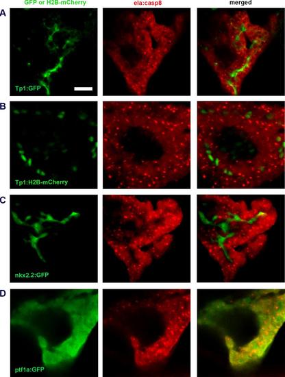
Confocal images of control animals ad. Fig. 4 Confocal images of untreated Tg(ela:casp8) at 11 dpf. A: Tg(Tp1:eGFP) labeling NRCs. B: Tg(Tp:H2B-mcherry) labeling NRCs in the nucleus.D: Tg(nkx2.2:eGFP) labeling ductal cells. E: Tg(ptf1a:eGFP) labeling ptf1a expressing cells. Scale bar: 20μm. |
Expression Data
Expression Detail
Antibody Labeling
Phenotype Data
Phenotype Detail
Acknowledgments
This image is the copyrighted work of the attributed author or publisher, and
ZFIN has permission only to display this image to its users.
Additional permissions should be obtained from the applicable author or publisher of the image.
Full text @ Dis. Model. Mech.