
Fig. 3
- ID
- ZDB-FIG-170315-4
- Publication
- Ziegler et al., 2017 - Thymosin β4 Improves Differentiation and Vascularization of EHTs.
- Other Figures
- All Figure Page
- Back to All Figure Page
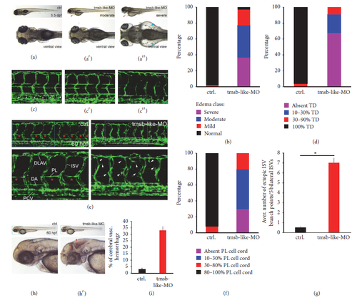
Effect of morpholino mediated tmsb-like knockdown in zebrafish. (a) Edema formation in zebrafish treated with tmsb-like-MO. Knockdown animals show moderate (a′) to severe (a′′) edema formation compared to control ((a), arrowhead), which was quantified in (b) (n=123 for control-MO and 114 for tmsb-like-MO). (c) Thoracic duct (TD) formation was blunted to a moderate (c′) to sever (c′′) degree in tmsb-like-MO animals compared to proper TD formation in control animals 5.5 days postfertilization (dpf, (c), arrowhead). Results were quantified in (d) (n = 112 for control-MO and 103 for tmsb-like-MO). (e) Knockdown of tmsb-like leads to the aberrant formation of intersegmental vessels (ISV) displaying an ineffective hypersprouting phenotype (arrowheads) and a perturbation of parachordal lymphatic vessel generation by parachordal lymphangioblasts (PL) 60 hpf. (PCV: posterior cardinal vein, DA: dorsal aorta, and DLAV: dorsal longitudinal anastomotic vessel). (f) Presence of parachordal lymphangioblasts in 10 successive somites (n = 109 for control-MO and 118 for tmsb-like-MO) and (g) average number of ectopic ISV branch points in 5 bilateral ISV (n=55 per group, *p < 0.05) demonstrate excessive branching and ineffective parachordal vessel formation in animals lacking tmsb-like. (h and i) tmsb-like-MO treated animals displayed a high rate of cerebral vascular hemorrhage compared to control-treated animals 60 hours postfertilization (n = 225 for control-MO and 204 for tmsb-like-MO, *p < 0.05). |