
Fig. 2
- ID
- ZDB-FIG-170227-24
- Publication
- Dalton et al., 2017 - Screening of anti-mycobacterial compounds in a naturally infected zebrafish larvae model
- Other Figures
- All Figure Page
- Back to All Figure Page
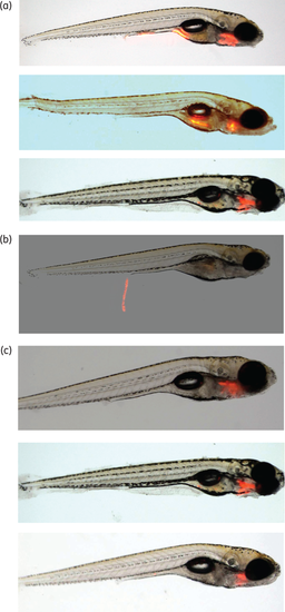
Natural exposure of zebrafish larvae to M. marinum results in transient colonization of the gut and infection of the developing gills and lower jaw. Larvae were exposed to M. marinum expressing a red fluorescent reporter and bacterial location identified by fluorescence microscopy. (a) Colonization of the developing gills and lower jaw 4 days post-infection. (b) Transient colonization of the digestive tract. (c) After 5 days, colonization is localized to the head region, with the digestive tract no longer colonized. Representative larvae are shown. This figure appears in colour in the online version of JAC and in black and white in the print version of JAC. |